
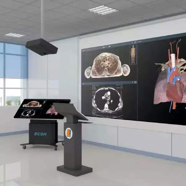
Medical students experience a steep learning curve when transitioning from textbook diagrams to live clinical settings. Understanding human anatomy requires profound spatial awareness. Traditional textbooks present flat, two-dimensional images. Physical specimens offer highly realistic anatomical references but lack repeatable modularity. Universities require instructional tools that actively build accurate three-dimensional cognition. Advanced virtual reality systems now fill this structural gap in medical curricula, providing a bridge between static observation and complex surgical navigation.
The Bottleneck of Traditional Spatial Training
Memorizing anatomical structures relies heavily on understanding adjacent relationships. Students must know exactly how a specific nerve cluster wraps around a corresponding vascular network. Achieving this level of comprehension in a traditional wet lab presents logistical hurdles. A physical specimen degrades with each use. Once a student dissects a specific tissue layer to view the underlying structure, that perspective is permanently altered.
This physical limitation prevents repeated practice. Medical departments face tight schedules and limited lab access. Students often get only one opportunity to view a specific anatomical region during an entire semester. Institutions require a supplementary platform that allows endless, non-destructive exploration of complex spatial relationships.
Tomographic Data Over Artistic Rendering
Commercial simulation software often relies on artistic 3D rendering. Artistic models may not fully represent the complexity required for some clinical education scenarios.
Artistic models lack the irregular nuances of real human tissue. Modern medical training requires actual, unedited human data. Advanced virtual platforms utilize continuous tomographic images sourced from human subjects with no organic diseases or physical defects.
These databases deliver uncompromising precision. The system utilizes two distinct datasets. The male dataset features 2,110 individual layers with a sectional precision between 0.1mm and 1mm. The female dataset provides 3,640 layers, pushing precision to 0.1mm to 0.5mm. By rendering these specific points, the software reconstructs over 6,000 distinct anatomical structures. The total pixel count across these tomographic images exceeds 1.2 billion. This guarantees that micro-vascular networks remain clearly identifiable even under extreme digital magnification.
Immersive Interaction and Spatial Tracking
Developers like DIGIHUMAN construct dedicated helmet-type stereo interactive systems to resolve spatial learning deficits. Users enter an immersive virtual environment built from digital anatomical datasets. Immersive hardware provides a wider field of view compared with traditional monitor-based visualization. Hand controllers allow students to grab, rotate, and strip away specific tissue layers from a first-person perspective.
Implementing human anatomy VR for institutions establishes a standardized environment for spatial training. The system utilizes six-degree-of-freedom tracking to mirror natural hand movements with high spatial precision. Instructors can monitor student actions in real-time. Students can trace the path of the sciatic nerve or isolate the entire cardiovascular system in seconds. If a learner makes an incorrect dissection during a practice module, the software resets the environment immediately. The VR space becomes a controlled virtual laboratory for repeated surgical planning.
Real-Time Processing for Lecture Flow
Processing 1.2 billion original pixels requires immense computing power. Standard commercial engines buffer or lag under this exact load. Educational environments cannot tolerate software delays during live instruction. A long loading time may interrupt classroom engagement.
Advanced platforms utilize proprietary processing architectures like the Tai engine. This specific framework guarantees loading speeds of under 30 seconds. A professor can jump directly from the central nervous system to the skeletal system without breaking the lecture’s flow. The user interface includes multi-mode displays and semantic association tools. Educators easily highlight target structures, hide surrounding tissue, and maintain flexible control over the pacing of the visual output.
Bridging Gross Anatomy and Diagnostic Imaging
Gross anatomy must connect directly to clinical diagnostics. A surgeon rarely views a fully exposed organ before an operation. They rely on radiological scans. Modern VR platforms merge 3D structural models with 2D hospital scans. The software integrates over 1,700 real CT and MRI images.
Students view the volumetric organ alongside its corresponding radiological scan. This split-screen approach trains the eye to identify pathological abnormalities across multiple diagnostic mediums. The system also includes human peel tools and see-through modes. Users observe the exact connection points between superficial layers and deep internal tissue, replicating the precise visual layers encountered during actual surgery.
Robust Assessment and Independent Study
Evaluating student progress with DIGIHUMAN requires robust digital frameworks. The virtual platform functions as a comprehensive digital repository for independent study. Assessment modules include self-testing modes with built-in anatomical term identification. The system supports multi-language switching, enabling international cohorts to study structures in English or regional medical terminology. Instructors gain access to a massive bank of digital practice questions. Students can review over 130 micro-class videos showcasing specific dissection animations. These integrated tools allow medical departments to track cohort knowledge retention continuously. Faculty can verify a student’s spatial comprehension before clearing them for actual clinical rotations.
High-fidelity virtual environments help overcome the spatial limitations of traditional educational mediums. Medical departments gain a repeatable, high-precision instructional tool. By integrating exact tomographic data, universities establish scalable, ultra-low risk laboratories that actively prepare cohorts for modern clinical environments.